

{{product.productLabel}} {{product.model}}
{{#if product.featureValues}}{{product.productPrice.formattedPrice}} {{#if product.productPrice.priceType === "PRICE_RANGE" }} - {{product.productPrice.formattedPriceMax}} {{/if}}
{{#each product.specData:i}}
{{name}}: {{value}}
{{#i!=(product.specData.length-1)}}
{{/end}}
{{/each}}
{{{product.idpText}}}
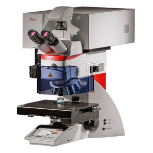
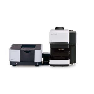
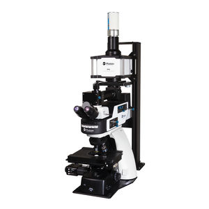
... you see in the microscope image - within a second. • - 2 systems in 1 for visual & chemical analysis • - 1 second to a chemical fingerprint • - 0 sample preparation Done! Examine exactly what you see via the eyepieces or camera ...
Leica Microsystems GmbH
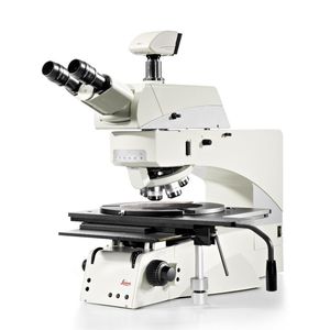
... illumination is based on the latest LED technology and is fully integrated into the microscope. The low heat radiation and integration into the stand ensures that there is an optimal airflow around the microscope. ...
Leica Microsystems GmbH

... lifetime of up to 25,000 hours. Further advantages: • - LED lights your samples homogeneously and at a constant color temperature, providing you with a realistic impression of your sample • - LED adjusts light intensity ...
Leica Microsystems GmbH
Magnification: 10 unit - 250,000 unit
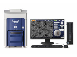
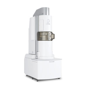
... sample stage is done using an automatically generated, zoomable colour photo overview. This can be supplemented locally by SEM photo snapshots. Product features: - EDX with 30mm2 or 60mm2 sensor size can be fully integrated, immediate ...
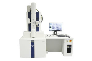
Magnification: 1,000,000, 800,000, 600,000 unit
Resolution: 0.2, 0.14, 0.19 nm
... Convenient on-screen operation via the fully integrated digital CMOS overview camera with >30 images per second instead of a bi-ocular - Electron source either tungsten or LaB - Fully integrated main cameras ...
Magnification: 2,000 unit
Resolution: 0.5, 0.1, 1 µm
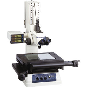
... The MF measuring microscope has a measuring accuracy that is highest in its class, and it follows the standards of JIS B 7153. It has a good expandability that makes it more reliable when associated with Mitutoyo's vision unit to enhance ...
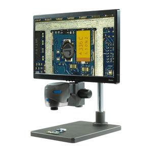
Magnification: 1 unit - 330 unit
Resolution: 10 nm
... requires liquid nitrogen.
Key Features
- Wide-field camera plus infrared microscope camera for observation areas up to 10 × 13 mm.
- 330× digital zoom enabling
Weight: 25 kg
Length: 29.5 cm - 29.5 cm
Width: 21.5 cm
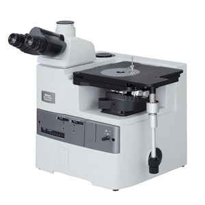
... Product overview
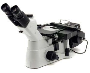
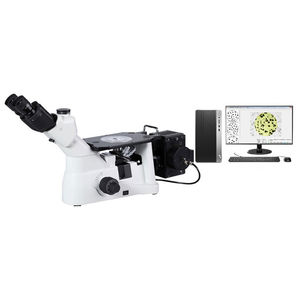
The ECLIPSE MA200
LED is an inverted metallurgical
microscope designed for episcopic optical-contrast inspection combined with
digital imaging accessories. It is intended for ...
Nikon Metrology
Resolution: 0.08 nm - 0.23 nm
... technique (e-ABF: enhanced ABF), facilitating observation of light-element materials, even at low accelerating voltages. The microscope room is separated from the operation room to respond to a remote operation. In addition, JEOL products’ ...
Jeol
Magnification: 32, 50, 6, 10 unit
... Compact full HD digital microscope system for quick and accurate inspection right where you need it. High quality custom-made optics and digital features combine for superior performance. Ideal for visual ...
Vision Engineering Ltd.
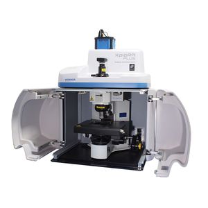
... The XploRA PLUS is a multi-user, multi-sample Raman microscope for research and analytical labs. Its fully confocal design guarantees quality images with the best spatial/depth resolution. Imaging with the SWIFT ™ is ten times faster ...
HORIBA Scientific

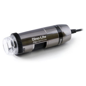
Magnification: 10 unit - 220 unit
Resolution: 650 nm
Weight: 137 g
... Field (EDOF)* • Extended Dynamic Range (EDR)* • Automatic Magnification Reading (AMR) • Adjustable polarizer • Flexible LED Control (FLC) ...
Dino-Lite Europe/IDCP BV
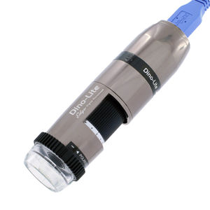
Magnification: 10 unit - 140 unit
Weight: 137 g
Length: 10.5 cm
... • Automatic Magnification Reading (AMR) • Adjustable polarizer • Flexible LED Control (FLC) Specification Lighting Light/ LED type - White Number of LEDs - 8 LED on/off switchable: - Yes Infrared ...
Dino-Lite Europe/IDCP BV
Magnification: 400 unit - 470 unit
Weight: 110 g
Height: 11.9 cm
... (EDR) • Automatic Magnification Reading (AMR) • Enhanced Flexible LED Control (eFLC) • Integrated polarizer (anti-reflection) • And more... Specification Lighting Light/ LED type - White Number of LEDs - 8 Polarizer ...
Dino-Lite Europe/IDCP BV
Magnification: 50 unit - 1,000 unit
Length: 25 cm
Width: 16 cm
... Product Introduction
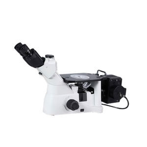
The WY-E inverted metallurgical
microscope is fitted with an infinity-corrected chromatic aberration correction optical system (CSIS), providing improved image quality and enhanced resolution for metallographic ...
Magnification: 50 unit - 500 unit
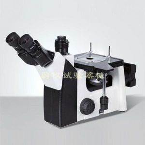
... It is a new technology in the field of microscope to input microscopic imaging into microcomputer and to process images by microprocessor. Image metallographic microscope, connected to a high-definition CCD camera ...
... A new design of infinite optical system, ergonomics can 360° rotary binocular observation tube, low-hand operation mode, to meet the user's long-term observation and use. A new ECO infrared sensing system with environmental protection and economic design ...
Magnification: 40 unit - 2,000 unit
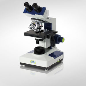
... Binocular and Trinocular Microscopes for Laboratory / Schools / Universities MBL2000 Series – Laboratory microscopes Robust and universal. This model is ideal for general microscopy in laboratories, schools and universities. ...
A. KRÜSS Optronic GmbH
Magnification: 40 unit - 800 unit
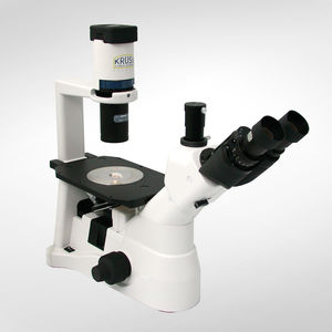
... petri dishes. With an upright microscope, the lens is under the specimen and the condenser above the specimen, which is particularly suitable for viewing on microscope slides. In an inverted microscope, ...
A. KRÜSS Optronic GmbH
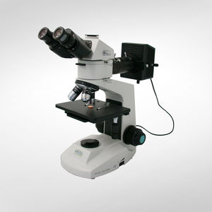
Magnification: 40 unit - 800 unit
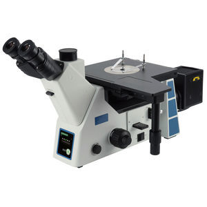
... structures following heat treatment. This metallurgical microscope is particularly well suited for laboratory and industrial applications. It is equipped with a phototube for connecting a camera. ...
A. KRÜSS Optronic GmbH
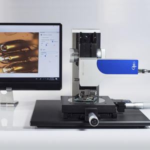
... The new "Machine Vision Microscope" (MVM) is a purely digital microscope with all the features that make a microscope. It has an apochromatically highly corrected microscope ...
Resolution: 200 nm
Weight: 1 kg
... USB/GigE digital microscope with optimized condenser and transmitted light LED. It fits to every lab and could be part of every thesis as it is programmable with Python and the budget needed is a fraction of that of ...
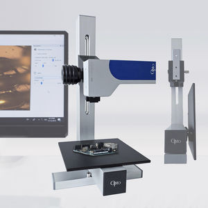
... The new ultra-compact digital Micro Measurement Microscope from Opto is the perfect tool for on-site measurements in production. The Machine Vision Microscope professionally digitizes quality where ...
... vertical movement and easy height adjustment, making it perfect for coin observation and welding. Easy digital microscope camera Easy digital microscope camera Capture ...
... Universal binocular microscope for precise, comfortable inspection The binocular microscope is suitable for all types of work requiring detailed observation and optimum comfort of use. It is supplied with a sturdy ...

Weight: 30 kg
Length: 1,000 mm
Width: 600 mm
... The microscope system PAMAS FastPatch 2 GO has been developed to comply with the standard ISO 4407 as an automated filter membrane particle counting and sizing system. The system is designed to measure the particles trapped on the surface ...
... analyzes. Entry-level inverted microscope for general purpose applications for hardness testing. Industrial and materials science inverted microscope especially designed for opaque specimens (including metals microstructure ...
Resolution: 2, 4 nm
Weight: 80 kg
Length: 150 cm
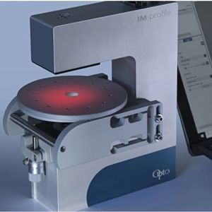
... Global hyperspectral microscope IMA is a hyperspectral microscope delivering spectral and spatial information in the VIS, NIR, and SWIR range (400 nm - 1620 nm). This system rapidly maps photoluminescence, electroluminescence, ...

Weight: 4 kg
Width: 200 mm
Height: 300 mm
... Study microscope. Standard model that has been equipped and transformed in order to study its optical characteristics. All dimensional parameters are measurable and adjustable. The study and understanding of the microscope ...
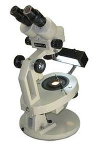
... gemological microscopes based on our versatile EM Series of stereo microscopes. Those who must work to the most demanding standards of the precious mineral industries will appreciate our full featured GEM Series. If you'd ...
MEIJI TECHNO
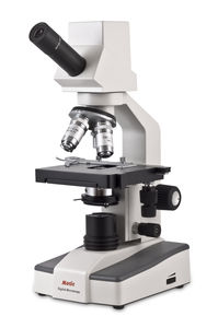
... of Digital Microscopy the DM-111 is the ultimate full scale full-scale professional unit equipped with all professional microscope features with added possibility of 100X oil microscopy. A 1.25NA Abbe condenser coupled ...
Motic
Magnification: 1 unit - 5 unit
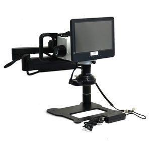
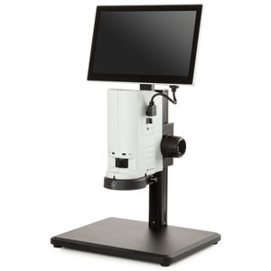
... MacroZoom MZ.5000 Digital The digital MacroZoom microscope is built around one 0.7 to 5x zoom objective and enables inspection of objects directly on an 11.6” LCD screen, -5° to 15° inclination. This ...
Magnification: 10, 4, 100, 40 unit
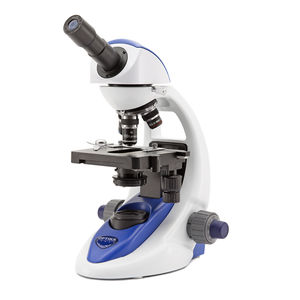
... yet sturdy and resistant, it is equipped with all the main controls to start learning how to use an advanced microscope and with long lasting LED illumination to provide over 20 years of use. Observation mode: Brightfield. Head: ...
Optika Srl
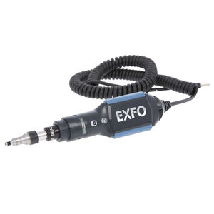
... Highly versatile probe to detect dirty/damaged connectors with unparalleled precision. Zoom In on Clarity It’s a known fact: optical network problems are often caused by dirty and/or damaged connectors. Using a fiber inspection probe to ensure that ...
Length: 677 mm
Width: 231 mm
Height: 384 mm
... material testing in a variety of industries. Observation tube Gemel trinocular, adjustable diopter, 45° inclined, with a digital camera port to shoot photo or video. Capture and save observation images. Image analysis ...
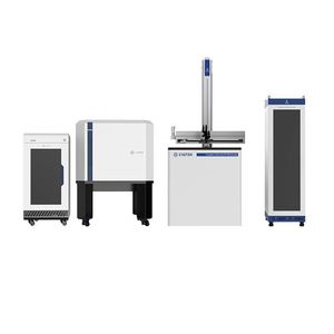
... The CIQTEK Scanning Nitrogen-vacancy Microscope (SNVM) is an advanced scientific analytical instrument that combines optically detected magnetic resonance (ODMR) of diamond nitrogen-vacancy centers with the scanning probe technique. This ...
CIQTEK Co., Ltd.
... distance objectives, endoscopic operation is extremely easy. 1.4. Binocular observation can be synchronized with photography, camera shooting. 1.5. With anti-static function, protect sensitive components from electrostatic damage. 1.6. ...
Shanghai Hualong Test Instruments Corporation
the best suppliers